Review of Rapid On-Site Evaluation (ROSE) Cytology Course at NEPSEC
Megan Drummond - Registered Biomedical Scientist, Newcastle Upon Tyne Hospitals NHS Foundation Trust
Introduction
I am a Histology based Biomedical Scientist who cross covers with Cytology. I started my career at North Tees University Hospital as a Healthcare Science Assistant in Histology in 2018 and after about a year, I moved into a Healthcare Science Associate role and began my registration portfolio. When the Cervical Cytology service was centralised our Cytology department lost many staff, I had the opportunity to train in Diagnostic Cytology. I became a HCPC registered Biomedical Scientist at the Royal Victoria Infirmary (RVI) in Newcastle in 2020.
When COVID restrictions started to lift in 2022 the Cytology department at the RVI re-introduced their Endobronchial Ultrasound Rapid On-Site Evaluation (EBUS ROSE) service and because of my previous experience in Cytology coupled with the fact the department wanted to encourage more rotation, I was offered the chance to train in ROSE. I started the initial training in the lab and was quickly taken to attend the EBUS clinic at the Freeman Hospital. I am now fully trained and after sign-off from our pathologist I now attend the EBUS clinic unaccompanied.
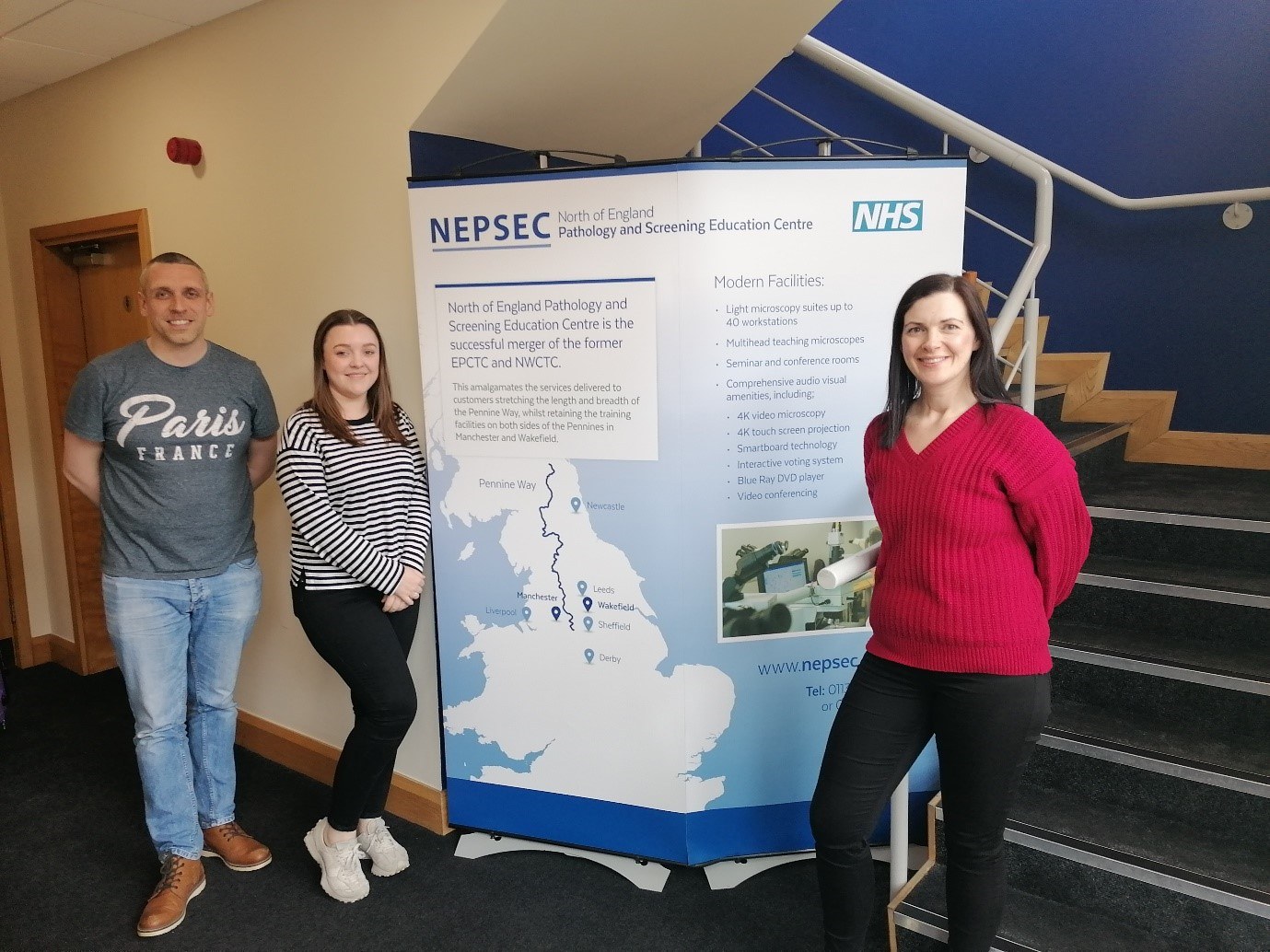
To further enhance my knowledge I attended the three-day North of England Pathology and Screening Education Centre’s ‘Rapid On-Site Evaluation of Cytology’ course in Wakefield. This was a new course, which aligns with the Institute of Biomedical Science (IBMS) new ROSE qualification, and my colleagues and I were excited to attend.
Day 1
On the first day we were introduced to our course tutors who were very friendly and welcoming along with the other course attendees. The other course attendees ranged from newly qualified BMS’s to Consultants, all of whom were actively attending ROSE clinics or reporting at them, and those looking to introduce a ROSE service in their department. Day 1 focused on head and neck FNA’s therefore we started with an introduction and the principals of ROSE in general. We discussed the benefits the service can bring along with the general technique. It was very interesting seeing a slightly different method and I definitely took away some ideas that we could incorporate in our current procedure and techniques.
We then moved on to head and neck pathology discussing thyroid, salivary gland and parotid glands along with some of the associated disease. We discussed the categorisation of thyroid masses from Thy1 - Thy5 and their microscopic appearances on FNA. We also looked at case studies from salivary glands FNA samples showing pleomorphic adenoma, Warthin's tumour, mucoepidermoid carcinomas and adenoid cystic carcinomas. We also touched on neck lymph node biopsies and Hodgkins's Lymphoma. All the while learning what an adequate or inadequate sample would look like for when we give our opinion during a ROSE clinic.
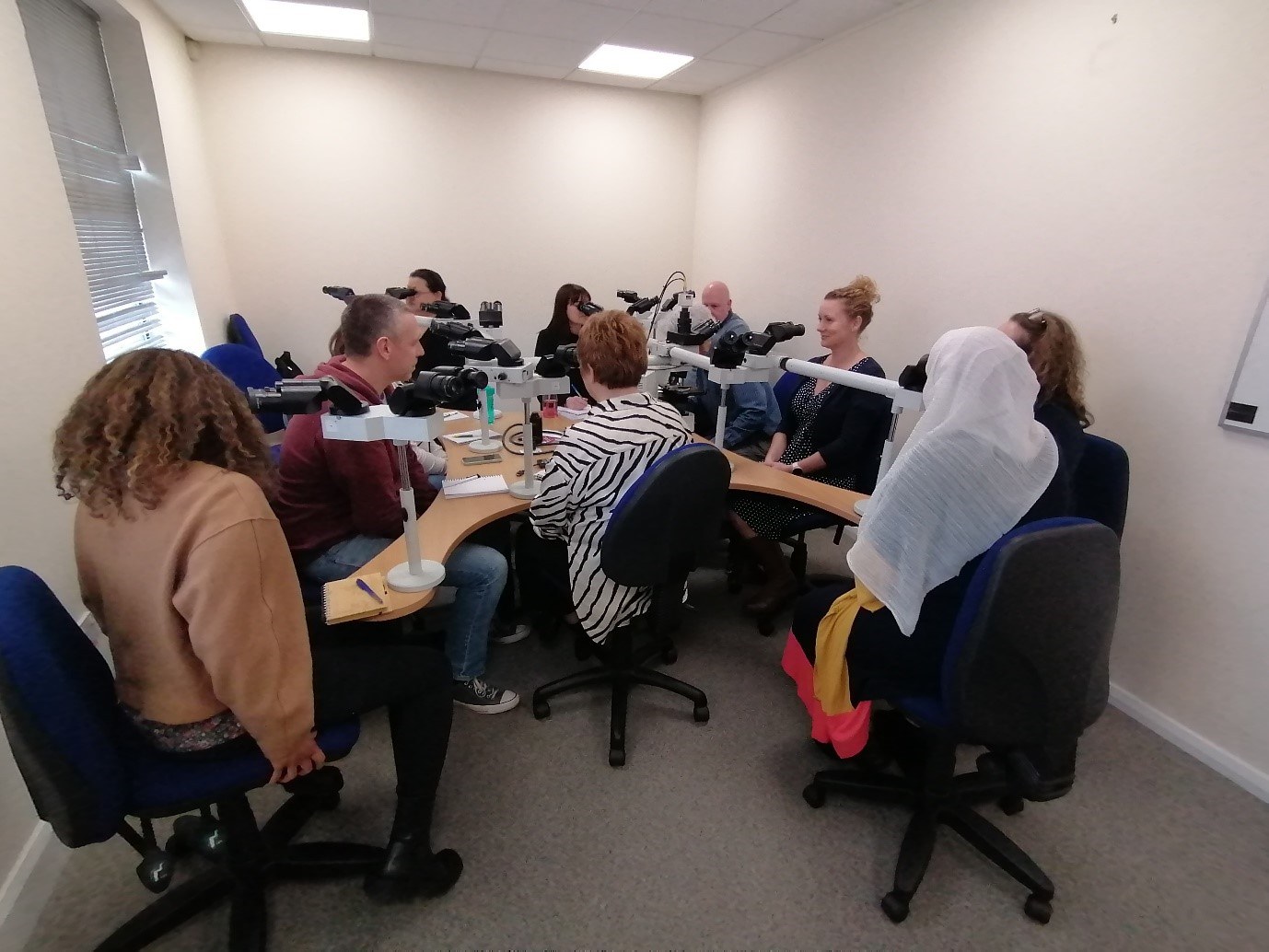
In the afternoon we had a fun practical activity which included looking at case slides under a microscope. We had 30 seconds to assess adequacy and another 30 seconds for those who had more experience to try to diagnose the sample. We then sat around the multi-header and discussed the cases. This was an interesting, fun activity and as someone who has no experience in head and neck FNA’s I used what I learnt about the different pathologies earlier in the day to attempt to assess the samples for adequacy and even took a chance at diagnosing some cases.
Day 2
On the second day we started with an introduction to EBUS, and the techniques used to obtain the sample and the method we ourselves use which was again very beneficial as I was able to note down some changes we can make to our own procedures. We learned about the lymph nodes around the lungs/bronchus and how our samples can help with staging and diagnosis. We also focused on how we give our verbal assessment to the clinician as the patient is only under sedation and can potentially still hear you, it is very important that we choose our words wisely when giving our findings, not to give false information to the patient or risk the patient misinterpreting anything they may hear.
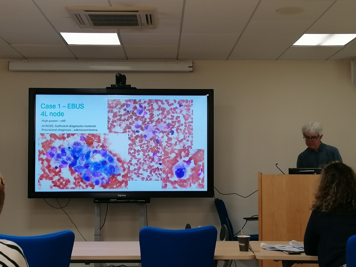
We then went on to the anatomy of the lungs and looked at a few examples of PET and CT scans that would be done before scheduling an EBUS procedure. We discussed different benign diseases such as sarcoidosis and granulomatosis and again how we determine adequacy within the samples in relation to the clinical details we are given. We then discussed a few case studies starting from clinical presentation, x-rays, CT scans, PET scans and EBUS, right through to Cytology and results, learning the different cell types and presentation of adenocarcinoma, squamous carcinoma, small cell carcinoma and high grade lymphoma.
In the second half of the day, we again had a fun practical on the microscope. Rapid-fire adequacy assessment for 30 seconds with an additional 30 seconds to attempt to diagnose the sample. With my previous knowledge and experience in EBUS I found this fun and engaging and even found myself getting the correct diagnoses on some of the cases which we discussed afterwards during a multi-header.
Day 3
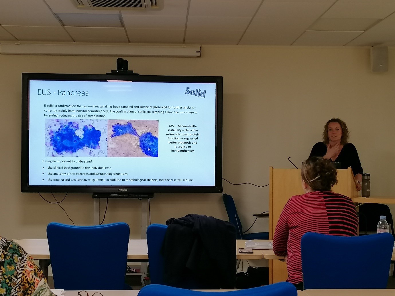
The third day was based on EUS biopsies of the pancreas which was all new to me. We had a brief overview about what EUS is and how the procedure is performed. I learned the risks of the procedure, the risks of multiple passes, and advantages of ROSE. We discussed solid tumours vs cysts and how sampling is a lot more complicated which makes assessing adequacy and ensuring there is enough sample for ancillary testing if needed of great importance. We discussed the anatomy of the pancreas, normal cytology and how lesions present for example pancreatic ductal adenocarcinomas and pseudocysts/simple cysts. We discussed the presentation and how to identify the cells of different tumours such as pancreatic ductal carcinomas, pancreatic NETs and Acinar Cell Carcinoma. We then discussed case studies from presentation to EUS to diagnosis. We then ended the day going through cases on the multi-header and discussing our opinions and diagnosis.
Conclusion
As a Histology based BMS the chance to learn something new in ROSE and being able to develop my skills has been very interesting and rewarding. Being trusted and confident enough to assess samples and to give my opinion makes me feel accomplished in my work and the ability to branch out in my career. The course was taught at a good level, as someone who has only attended EBUS it was great to learn about the other types of clinics we can eventually expand our services to and learn new pathologies along the way. Learning about how other laboratories use different techniques allows me to take different methods and ideas that could be adapted to our current practice to potentially improve our service and therefore patient results. The course was fun and informative, and the tutors were friendly, approachable and no question went unanswered. I would highly recommend this course if you would like to improve your knowledge of ROSE, just starting out or are thinking about setting up your own ROSE service within your Trust. Obtaining an adequate sample improves the patient’s results and experience and therefore puts the patient at the heart of the service.